 CirascanTM is a specially designed imager that captures multiple images from a Ciraplex® array, using an astronomy-grade cooled CCD camera with high-end optics. The imager uses a patent pending multiple exposure image acquisition algorithm to produce a high resolution composite image of each chemiluminescent array plate. An easy to use interface allows new users to operate Cirascan with minimal training. Simply prep samples using familiar ELISA protocols, place the plate into the compact, benchtop imager, and receive fully analyzed results within minutes!
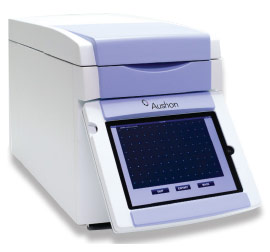
Cirascan' s intuitive Apple iPad touch screen user interface allows researchers to select a plate layout created at their desktop computer and enter it on Cirascan via a network connection, Sample identification information can be entered directly into a plate layout with the optional CiraTM barcode reader.
CirascanTM is designed to capture a series of images, offering an improved broader dynamic range of assay quantification. Rapid imaging (typically <2 minutes per plate) and a fixed-focus setting 每 so no focus adjustments are required from one image to the next 每 make it easily shared by multiple users. Plus, automated calculations of image time make the instrument incredibly easy to use.
| Complete System | Imager (integrated with embedded Mac mini computer and iPadTM user interface), and array analysis software |
| Performance | Improved sensitivity and dynamic range |
| Fast | Typically generates 16-bit images in <2 minutes |
| User-friendly | Integrated touch screen user interface for image capture and storage; Fixed-focus setting and automatic calculation of optimal image times |
| Accuracy | Customized array software locates each spot in the image, measures density values and calculates pg/ml values for unknown samples from standard curves. Spot location and analysis is automatic and rapid. Providing nearly immediate access to measured sample concentrations. |
| Quality | Custom, high resolution camera/lens for improved image quality and more consistent results |
| Productivity | Image acquisition can take place at the bench, analysis at desk 每 freeing the imager for other users or to run more plates. |
| Secure | Images can be archived for a permanent record of raw experimental data |
| Set-up | Simple, fast, out-of-the-box installation 每 ready to use with minimal instruction |
| Compact | Uses <2 sq.ft. of bench space; All components integrated with embedded MacMini and iPad user interface, eliminating need for separate system components |
| Maintenance-free | Unlike fluidic array analysis systems, no daily, weekly or monthly maintenance is required |
| Dimensions | 11" W x 14 3/4" H x 24" D |
| Type of CCD | Front-illuminated |
| Single Exposure Data Acquisition | 16bit |
| Resolution | 2184 x 1472 pixels |
| Pixel Size | 6.8 x 6.8 米m |
| Single Exposure Dynamic Range | 65,000 grayscale |
| Cooling | -23∼C |
| Electrical Configuration | 100-240 VAC/1.8 amperes/50-60 Hz |